fiberoptic Bronchoscopy
- Home
- Services
- Pulmonology
- fiberoptic Bronchoscopy
What is Fiberoptic Bronchoscopy?
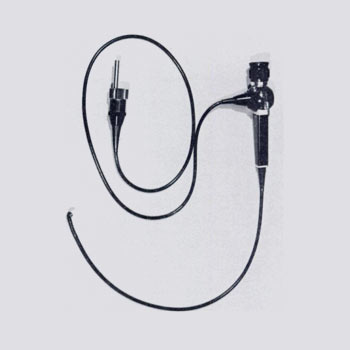
Fiberoptic bronchoscopy is a crucial diagnostic and therapeutic procedure in pulmonology, providing valuable insights into the respiratory system's health and aiding in various treatments. This advanced medical technique utilizes a flexible fiberoptic bronchoscope, a thin, flexible instrument equipped with a light source and a camera. It allows pulmonologists to visualize the inside of the airways and lungs in detail, facilitating both diagnostic examinations and therapeutic interventions
What are the main components of a Fiberoptic Bronchoscope?
A fiberoptic bronchoscope consists of several essential components
Flexible Shaft
The bronchoscope's flexible shaft, usually around 5-6 mm in diameter, contains bundles of optical fibers. These fibers transmit light from the source and images captured by the camera at the tip of the bronchoscope
Light Source
Positioned at the proximal end of the bronchoscope, the light source provides illumination through the fiberoptic bundles, enabling clear visualization of the bronchial tree
Camera and Lens System
At the distal end of the bronchoscope, a miniature camera and lens system capture high-resolution images of the airways and transmit them to a monitor for real-time viewing.
Channel(s)
Some bronchoscopes feature additional channels for inserting instruments such as biopsy forceps, brushes, or suction devices, enabling simultaneous diagnostic sampling and therapeutic procedures

What does the procedure of a Fiberoptic Bronchoscope involve?
Fiberoptic bronchoscopy procedures are typically performed in a specialized suite within a hospital or outpatient setting. The patient is usually sedated or given local anesthesia to minimize discomfort. The procedure involves
Insertion
The bronchoscope is inserted through the patient's mouth or nose and guided down the throat into the trachea and bronchial tubes
Visualization
As the bronchoscope advances, the camera captures detailed images of the airway walls, allowing the pulmonologist to inspect for abnormalities such as tumors, inflammation, or infections
Diagnostic Sampling
During the procedure, the pulmonologist may collect tissue samples (biopsies), washings, or brushings from suspicious areas for laboratory analysis, aiding in the diagnosis of conditions like lung cancer, tuberculosis, or pneumonia
Therapeutic Interventions
Fiberoptic bronchoscopy also facilitates therapeutic interventions such as the removal of foreign bodies, management of bleeding lesions, or the placement of stents to open blocked airways
What are the indications for Fiberoptic Bronchoscopy?
- Lung Cancer Diagnosis and Staging: By obtaining tissue samples for biopsy and assessing the extent of tumor spread
- Evaluation of Persistent Cough or Hemoptysis: To identify the underlying cause, such as bronchitis, bronchiectasis, or pulmonary embolism
- Evaluation of Airway Lesions: Including benign or malignant tumors, strictures, or foreign body aspirations
- Assessment of Infections: Including tuberculosis, fungal infections, or pneumonias where conventional diagnostic methods are inconclusive
- Therapeutic Interventions: To alleviate symptoms or improve respiratory function, such as removing secretions or placing stents
Benefits
Fiberoptic bronchoscopy is minimally invasive, providing direct visualization and precise diagnostic capabilities. It allows for targeted treatment and management decisions, improving patient outcomes
fiberoptic bronchoscopy is a versatile tool in pulmonology, offering comprehensive diagnostic insights and therapeutic interventions for a wide range of respiratory conditions. Its ability to combine visualization with minimally invasive procedures underscores its importance in modern pulmonary medicine, facilitating accurate diagnoses and improving patient care outcomes. As technology continues to advance, fiberoptic bronchoscopy remains pivotal in the ongoing effort to enhance respiratory health and quality of life.
