CVC Placement
- Home
- Services
- Pulmonology
- CVC Placement
What is CVC Placement?
Central Venous Catheter (CVC) placement is a crucial procedure in pulmonology and critical care medicine, involving the insertion of a catheter into a large vein for various medical purposes. Typically, CVCs are placed in veins near the heart, such as the internal jugular vein, subclavian vein, or femoral vein. This procedure is performed by trained medical professionals under sterile conditions to minimize the risk of infection and ensure accurate placement
What is the significance of CVC placement?
Central Venous Catheters serve several essential functions in pulmonary and critical care settings
Monitoring and Administration
CVCs allow for the continuous monitoring of central venous pressure (CVP), which is vital in managing fluid status and assessing cardiac function. They also facilitate the administration of medications, fluids, blood products, and nutrition directly into the central circulation.
Long-term Access
Unlike peripheral intravenous lines, CVCs provide long-term access for medications and treatments that cannot be safely administered through peripheral veins. This is particularly beneficial for patients requiring prolonged intensive care or chemotherapy
Emergency Access
In emergency situations, CVC placement enables rapid fluid resuscitation and medication administration, crucial for stabilizing critically ill patients

What are the different types of Central Venous Catheters?
There are several types of CVCs, each with specific indications and considerations
Single-Lumen CVC
A single-channel catheter used for basic fluid administration and monitoring.
Double-Lumen CVC
Provides two lumens, allowing for simultaneous infusion of medications and fluids, or blood sampling without disrupting infusion
Triple-Lumen CVC
Offers three lumens for complex medication regimens, frequent blood sampling, and continuous hemodynamic monitoring
How is CVC Placement performed?
The procedure for placing a CVC involves the following steps
Patient Preparation
The patient is positioned appropriately, usually lying flat with the head turned away from the insertion site. The area is cleaned and draped in a sterile fashion
Vein Selection
The clinician selects the appropriate vein for catheter insertion, often guided by ultrasound to ensure accuracy and reduce complications
Catheter Insertion
A needle is used to puncture the skin and vein, through which a guide wire is threaded into the vein. The catheter is then advanced over the wire into the vein until it reaches the desired position near the heart.
Confirmation and Securing
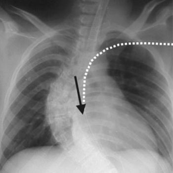
Placement is confirmed via imaging (e.g., chest X-ray or ultrasound) to ensure the catheter tip is correctly positioned. The catheter is then secured to prevent accidental displacement
Post-Procedure Care
Ongoing monitoring for complications such as bleeding, infection, pneumothorax (if subclavian vein accessed), and thrombosis is essential. Proper dressing and care of the insertion site are crucial to prevent infections
Complications and Considerations
Despite its benefits, CVC placement carries risks, including infection, thrombosis, and mechanical complications (e.g., catheter malposition). Careful patient selection, proper technique, and adherence to sterile protocols are critical in minimizing these risks
Central Venous Catheter placement is a fundamental procedure in pulmonology and critical care, enabling accurate monitoring, administration of medications, and long-term vascular access. Understanding the procedure, its indications, and potential complications is essential for healthcare providers managing critically ill patients
